








Cartilage
The smooth tissue covering the joint surfaces. Arthrosamid does not replace missing cartilage or grow a new joint surface.
A closer look at your treatment
A small injection. A different part of the joint.
Arthrosamid is a non-absorbable hydrogel that becomes incorporated into the knee’s inner lining. Understanding that lining helps explain what the treatment is designed to do.
01 / Find the right tissue
Cartilage covers the ends of the bones. The synovium lines the inside of the capsule around the joint. These are different structures, with different roles.
The smooth tissue covering the joint surfaces. Arthrosamid does not replace missing cartilage or grow a new joint surface.
The focus of this treatment
The living tissue inside the joint capsule. Arthrosamid becomes incorporated into this tissue after injection.
The space inside the capsule, containing synovial fluid. This is where the injection is delivered.
Where does this fit into your treatment options?
Start your suitability assessment02 / From injection to integration
The injection site and the eventual location of the hydrogel are different parts of the story.
The clinician places the hydrogel into the joint using ultrasound guidance. It is not injected directly into the synovial tissue or the cartilage.
Arthrosamid is already a hydrogel when it is injected. This is not a liquid being used to set into a hard replacement joint surface.
See what happens on treatment day →Histological studies have shown hydrogel becoming incorporated beneath the synovial surface, with connective tissue growing through it.
The illustration shows that relationship: hydrogel within the lining, rather than a new layer of cartilage between the bones.
This simplified tissue view is informed by animal histology. It does not show a precise human timetable or prove how pain relief occurs. Read the source notes.
The treatment is intended for symptom management. It does not reverse osteoarthritis or rebuild the worn joint surfaces.
Understand the mechanism. Then consider your own knee.
Start your suitability assessment03 / What this means for you
Published clinical research has reported improvements in pain and knee function after treatment. Your response depends on your individual clinical situation; a diagram cannot predict your outcome.
Explore the research and results →The synovium is part of the joint’s response to osteoarthritis. Researchers are studying how hydrogel integration interacts with the tissue and its cells. The complete pathway from integration to symptom relief is not fully established.
Arthrosamid is non-absorbable. The material remaining in the tissue does not mean that pain relief is permanent.
Our linked observational cohort followed outcomes over 24 months. That follow-up period is not an expiry date, a guarantee of two years’ relief or a prediction for your knee.
What could this mean for your symptoms and goals?
Start your suitability assessment04 / The person behind the treatment
Sharron Davies MBE shares her experience of Arthrosamid treatment with Professor Paul Lee.
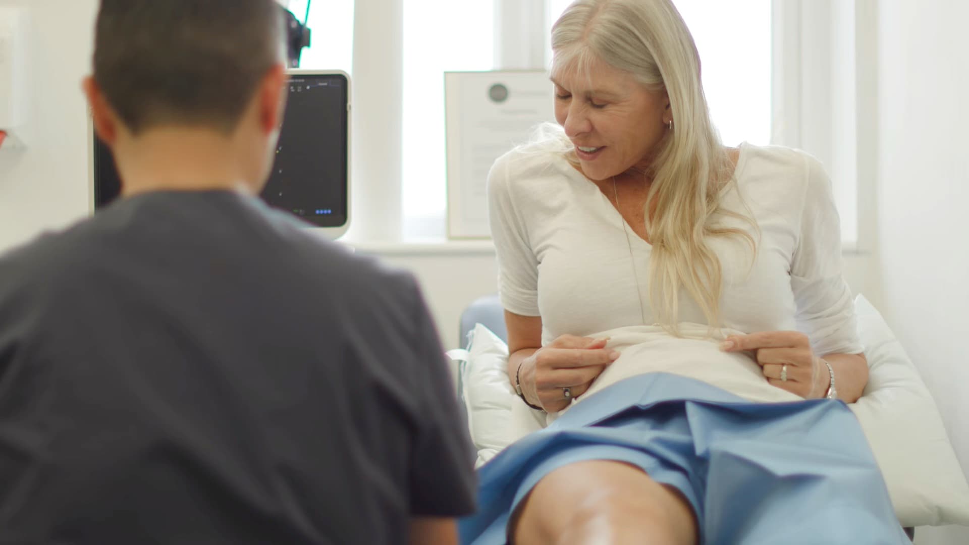 Watch Sharron’s story · 0:36
Patient perspective
A personal account can help you understand what it is like to choose treatment. It sits alongside the research and your own assessment.
One person’s experience. Results and recovery vary.
Start your suitability assessmentExplore patient stories →
Professor Paul Lee and the team assess your history, scans and previous treatment before discussing whether Arthrosamid belongs in your plan.
Arrange a discovery call →Look behind the illustrations
Animal histology supports incorporation into synovial tissue. It does not establish an individual human response or all proposed mechanisms of pain relief.
Product information on material properties and administration. Manufacturer information is distinct from independent clinical research.
Published results, study design and limitations, including the 24-month observational cohort.
Original explanatory illustrations. Simplified anatomy, not to scale. This draft deliberately separates demonstrated tissue integration from proposed cellular explanations.
Your next step
The questionnaire helps prepare for a consultation. It cannot diagnose your condition or confirm suitability.